Overview
Machine learning (ML) techniques are making progress in simplifying the detection of damage caused by myocardial infarction (MI) using cardiac magnetic resonance (CMR) scans. Researchers part-funded by HDR UK believe their work is paving the way for new approaches to detecting scarred heart tissue more quickly, cheaply, and without the use of contrast agents.
The Challenge
Cardiovascular diseases (CVDs) caused approximately 18.6 million deaths (31% of all deaths) in 2022 according to the World Heart Federation. Many of these resulted from MI – myocardial cell death which can lead to the loss of contraction of the damaged portion of the heart muscle.
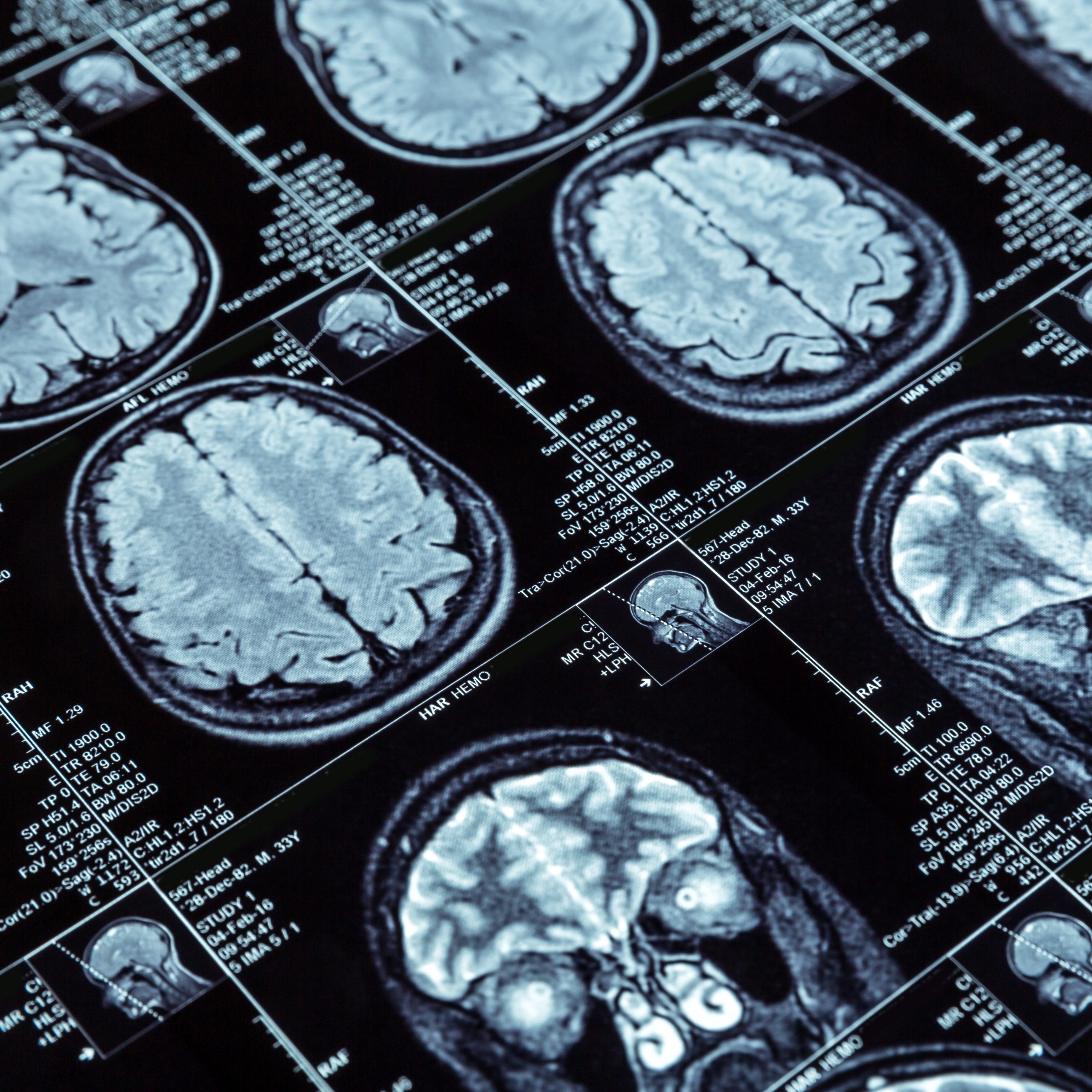
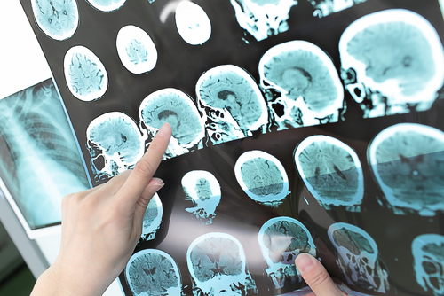
CMR imaging is effective for diagnosing MI, showing the presence, location and extent of scarring on the heart tissue. The patient requires an initial scan, after which contrast agents are administered and a second scan performed. The scans are then compared and used for diagnosis.
The research team set out to see if ML techniques could be used to analyse the first scan and accurately predict what the second would show. This could lead to quicker scans, simpler diagnosis, shorter appointments and lower costs. It would also extend the use of CMR to patients, such as those with severe kidney disease, for whom contrast agents are unsafe.
Solution
The research team (which was international and involved a wide variety of leading organisations and institutions) studied 272 retrospectively selected CMR studies, 108 showing MI and 164 healthy controls. They applied deep learning segmentation and classification models to discover the extent and location of any scars. With radiomics approach, complex data patterns were captured and used to try to develop predictive models that would allow pre-contrast scans to predict what would be found using post-contrast scans.
Impact
The findings were published in a paper entitled Predicting post-contrast information from contrast agent free cardiac MRI using machine learning: Challenges and methods (published in Frontiers in Cardiovascular Medicine, July 2022).
The work represents an original contribution to the area and suggests that sufficient information could be available from pre-contrast CMR scans to identify MI damage.
The research presents new parameters (such as rate of myocardial area change, optical flow and radiomics parameters) that could be considered biomarkers of the mechanics of myocardial disease.
Overall the team successfully developed promising techniques that, with further work (and, specifically, much larger datasets and more advanced ML techniques), could lead to practical and inexpensive methods for using ML to diagnose MI without the use of a contrast agent.
Lead author Dr Musa Abdulkareem, a Principal Research Healthcare Scientist at Barts Health NHS Trust, said: “The results are not 100% perfect at this stage. But from what we have done we can see that the information contained in the pre-contrast images can, in future, allow us to predict what can be seen using post-contrast images.
“If you solve this problem, you will profoundly affect cardiovascular imaging.”
What the Impact Committee said:
The committee praised the study for its contribution to applied analytics and as an example of strong collaborative research through international and multi-institutional collaboration.
Contact